Uncategorized

This Is What a Donated Human Body Looks Like After Science Gets It – Mind-Blowing Slices
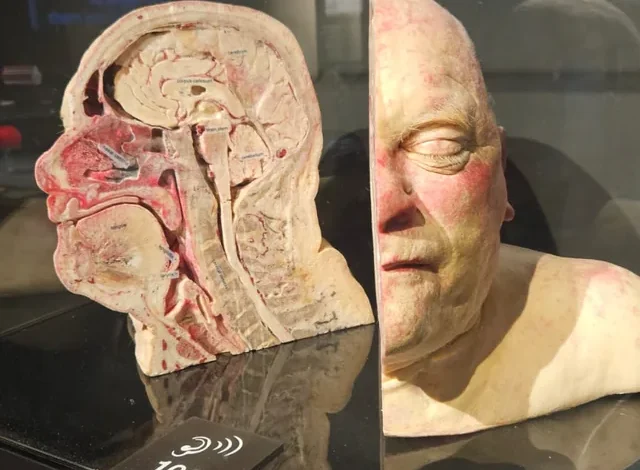
This striking image shows a preserved human specimen meticulously sectioned into thin layers—a powerful example of the kind of anatomical material made possible through voluntary body donations to science.Body donation is a deeply generous, entirely consensual act: during their lifetime, individuals explicitly authorize that after death, their remains be used for medical education, research, and training. These gifts allow future doctors, surgeons, anatomists, and health professionals to work with real human tissue rather than models or simulations.
Such donations enable hands-on study of authentic human anatomy in unprecedented detail. Specialists can explore complex structures, rehearse intricate surgical procedures, examine pathological changes in diseased organs, and deepen their understanding of how the body functions in health and illness. Preparing these specimens often involves advanced preservation techniques—like plastination (a process invented by Dr. Gunther von Hagens that replaces bodily fluids with durable polymers) or precise cross-sectioning (freezing the body, embedding it, and slicing it into ultra-thin sheets)—to create long-lasting, odor-free, and anatomically accurate teaching tools.While the sight of sliced or dissected bodies can feel unsettling or even shocking at first glance, these are never random acts of mutilation. Every cut, preservation step, and display is carefully planned, ethically regulated, and carried out with profound respect for the donor’s final wish: to contribute meaningfully to advancing medical knowledge.
The result? These transformed specimens become extraordinary educational resources—helping train generations of healthcare providers, improve surgical skills, accelerate disease research, and ultimately save lives. What might appear macabre is, in reality, one of the most selfless contributions a person can make to humanity’s progress in medicine and science.